Capnography is one of the most clinically dense monitoring tools available in the ICU — and one of the most underutilized. For the registered nurse working in a critical care environment, understanding end-tidal carbon dioxide (EtCO₂) monitoring goes far beyond confirming endotracheal tube placement. It provides real-time insight into a patient’s ventilation, perfusion, metabolism, and even cardiac output. Whether preparing for the NCLEX or working a night shift in a medical ICU, mastering capnography EtCO₂ ICU nursing principles is non-negotiable for safe, evidence-based practice. This guide — drawn from the same foundations as the rn-nurse.com nursing bundle — breaks down the full clinical picture.
What Is Capnography and How Does EtCO₂ Work?
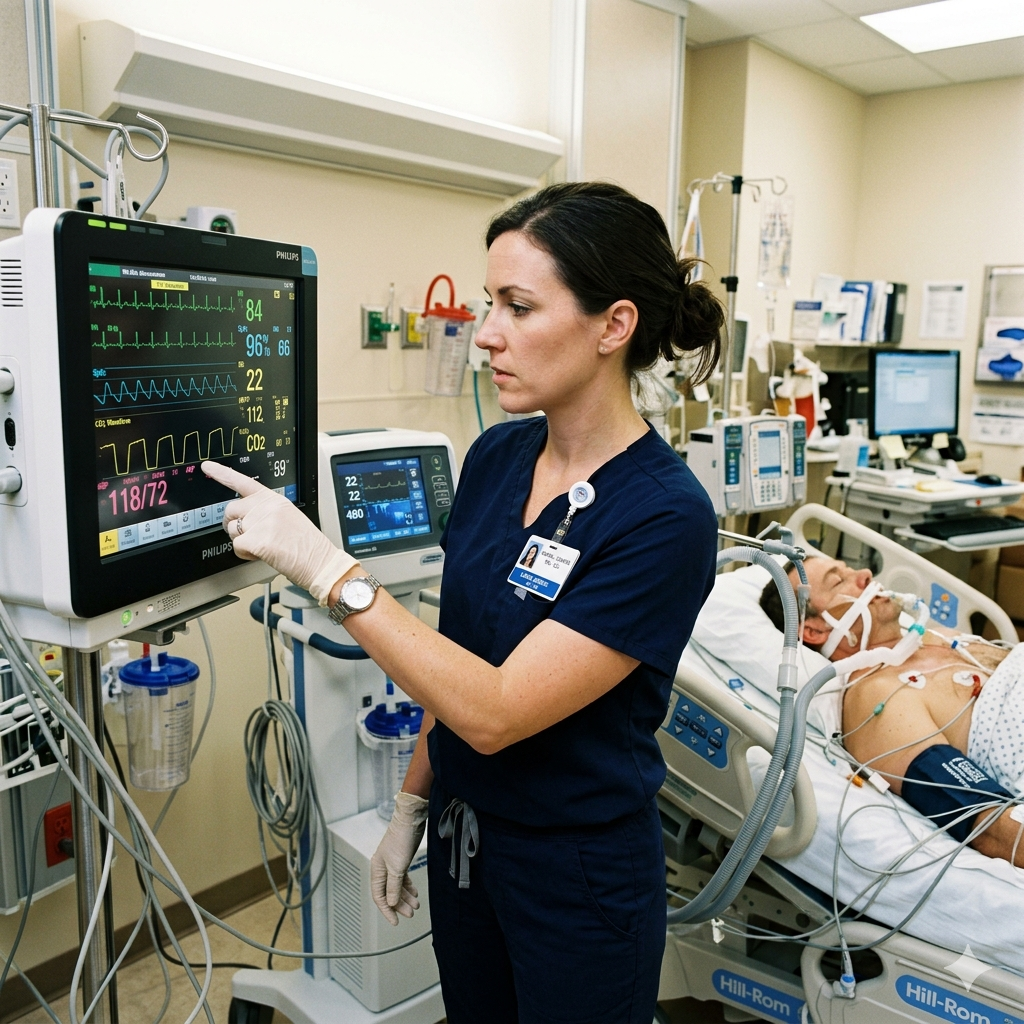
Capnography is the continuous, non-invasive measurement of carbon dioxide (CO₂) in exhaled breath. The primary value reported is EtCO₂ (end-tidal CO₂), which reflects the partial pressure of CO₂ at the end of a full exhalation. Normal EtCO₂ is 35–45 mmHg, closely mirroring arterial CO₂ (PaCO₂) with a gradient of approximately 2–5 mmHg.
The capnograph produces a waveform — the capnogram — that traces the CO₂ concentration across the respiratory cycle. This waveform has four distinct phases:
- Phase I (A–B): Baseline — dead space exhalation, CO₂ ≈ 0
- Phase II (B–C): Expiratory upstroke — alveolar gas begins mixing with dead space
- Phase III (C–D): Alveolar plateau — peak CO₂ concentration; EtCO₂ is measured at point D
- Phase IV (D–E): Inspiratory downstroke — CO₂ returns to zero as fresh gas enters
A normal capnogram has a rectangular appearance with a flat alveolar plateau. Deviations from this shape are clinically significant and guide nursing assessment and intervention.
Capnography EtCO₂ ICU Nursing: What the Numbers Really Tell You
The RN nurse must understand that EtCO₂ reflects three integrated physiological processes: ventilation, perfusion, and metabolism. Each component can independently alter EtCO₂ readings.
| Parameter | Effect on EtCO₂ | Clinical Cause |
|---|---|---|
| ↑ Ventilation | ↓ EtCO₂ | Hyperventilation, anxiety, pain |
| ↓ Ventilation | ↑ EtCO₂ | Hypoventilation, sedation, airway obstruction |
| ↑ Perfusion/CO | ↑ EtCO₂ | Fever, sepsis (early), increased metabolism |
| ↓ Perfusion/CO | ↓ EtCO₂ | Cardiac arrest, PE, hemorrhagic shock |
| ↑ Metabolism | ↑ EtCO₂ | Malignant hyperthermia, thyroid storm |
| ↓ Metabolism | ↓ EtCO₂ | Hypothermia, decreased CO |
This interplay is exactly why nursing assessment must always correlate EtCO₂ with clinical context — a falling EtCO₂ can indicate overventilation or impending cardiovascular collapse.
Waveform Analysis: What Every ICU Nurse Must Recognize
Skilled capnography interpretation requires the registered nurse to read beyond the number and analyze the waveform shape.
Shark fin waveform (obstructive pattern): The alveolar plateau rises in a sloped, shark-fin appearance rather than a flat shelf. This is the classic sign of bronchospasm or COPD exacerbation. The expiratory upstroke is prolonged due to uneven airway emptying. Nursing action: assess for wheezing, notify provider, prepare bronchodilator therapy.
Elevated baseline: If CO₂ does not return to zero during inspiration, consider rebreathing — a malfunctioning CO₂ absorber, faulty expiratory valve, or an exhausted HME filter.
Sudden drop to zero: An abrupt flatline capnogram is a critical nursing alert. Causes include accidental extubation, circuit disconnect, complete airway obstruction, or cardiac arrest. Immediate assessment and intervention are required.
Cleft in plateau (cardiogenic oscillations): Small notches on the alveolar plateau may reflect the cardiac cycle impressing on ventilation — often seen in low tidal volume or high heart rate scenarios. This is benign but worth recognizing on NCLEX.
EtCO₂ in Cardiac Arrest: A Perfusion Marker
Perhaps the most clinically impactful application beyond ventilation is EtCO₂ use during cardiopulmonary resuscitation (CPR). During cardiac arrest, CO₂ accumulates in tissues and venous blood. As CPR generates cardiac output, CO₂ is transported to the lungs and exhaled — making EtCO₂ a real-time surrogate for cardiac output and CPR quality.
Key NCLEX and clinical principles:
- EtCO₂ < 10 mmHg during CPR despite adequate compressions suggests poor cardiac output and a poor prognosis
- EtCO₂ ≥ 10–20 mmHg indicates effective chest compressions are generating meaningful perfusion
- A sudden, sustained rise in EtCO₂ (often to 35–40 mmHg) during CPR is a strong indicator of return of spontaneous circulation (ROSC) — frequently detected on capnography before a palpable pulse is confirmed
This makes capnography an essential tool during code situations. The RN nurse must recognize that a rising EtCO₂ during resuscitation is not a ventilator artifact — it is a sign the heart may be resuming function.
Procedural and Non-Intubated Applications in the ICU
Capnography EtCO₂ ICU nursing extends beyond the ventilated patient. Many ICUs now utilize sidestream capnography for spontaneously breathing patients via nasal cannula or mask adapters. Clinical applications include:
- Procedural sedation monitoring: EtCO₂ detects respiratory depression before SpO₂ drops — providing earlier warning of hypoventilation during bronchoscopy, intubation, or bedside procedures
- Post-extubation surveillance: A rising EtCO₂ following extubation may indicate fatigue, partial obstruction, or impending respiratory failure before clinical signs manifest
- Opioid-induced respiratory depression: ICU patients on PCA or continuous opioid infusions benefit from EtCO₂ surveillance as an early safety net
- Nasogastric tube confirmation: While not replacing radiographic confirmation, the absence of CO₂ waveform during tube insertion helps rule out inadvertent tracheal placement
Each of these applications demands nursing vigilance, clinical reasoning, and prompt escalation when values trend outside normal parameters.
💡 NCLEX Tips for Capnography EtCO₂
- Normal EtCO₂ = 35–45 mmHg; values outside this range require nursing assessment and correlation with clinical findings
- A sudden drop in EtCO₂ to zero = accidental extubation or circuit disconnect until proven otherwise — assess airway immediately
- During CPR, a sudden rise in EtCO₂ is the earliest sign of ROSC — do not stop compressions to check pulse without this context
- A shark-fin waveform = bronchospasm or COPD — prepare for bronchodilator administration
- EtCO₂ monitors ventilation and perfusion — a falling reading in a hemodynamically unstable patient suggests decreasing cardiac output, not just hypoventilation
Nursing Interventions and Documentation Priorities
The registered nurse caring for a capnography-monitored patient carries specific responsibilities that go beyond observing the number on the screen.
Establish baseline: Document EtCO₂ on initiation of monitoring and following any ventilator changes, sedation adjustments, or clinical status changes.
Correlate with ABGs: EtCO₂ approximates PaCO₂ but does not replace arterial blood gas analysis. When the EtCO₂–PaCO₂ gradient widens, suspect increased dead space ventilation — a marker of pulmonary embolism, decreased cardiac output, or V/Q mismatch.
Set alarm parameters: ICU nursing protocols typically alarm for EtCO₂ < 30 or > 50 mmHg. Nurses must ensure alarms are active, audible, and responded to promptly.
Troubleshoot sensor placement: Sidestream sensors are prone to moisture condensation and sampling line occlusion. The RN nurse should inspect connections regularly and replace sampling lines per unit protocol.
These documentation and surveillance skills are heavily tested in the nursing bundle review modules and appear frequently on NCLEX critical care question sets.
Conclusion
Capnography EtCO₂ ICU nursing is a high-yield competency that bridges ventilator management, hemodynamic monitoring, and resuscitation science. The RN nurse who can interpret waveforms, recognize perfusion changes, and act on real-time CO₂ data is better equipped to manage the complex, rapidly changing ICU patient. From detecting ROSC during a code to identifying bronchospasm on a capnogram, this skill set separates competent from expert critical care nursing practice.
Deepen your NCLEX readiness with targeted critical care practice questions at rn-nurse.com/nclex-qcm/, or explore the full critical care nursing bundle at rn-nurse.com/nursing-courses/ to master ICU monitoring from ventilators to hemodynamics.