A ventricular aneurysm after MI is one of the most serious late complications a nurse can encounter on a cardiac unit, and it is a favorite topic on the NCLEX because it tests both pathophysiology knowledge and priority-setting skills. When myocardial tissue dies during a heart attack, the scarred, weakened wall can begin to bulge outward under normal ventricular pressure, creating a thin, non-contractile pouch. For any registered nurse working in cardiac care, recognizing the early warning signs of this complication can mean the difference between a stable recovery and a life-threatening event such as rupture, embolism, or heart failure. Understanding this condition thoroughly is essential preparation for both clinical practice and exam success.
What Is a Ventricular Aneurysm?
A ventricular aneurysm develops when a segment of the myocardial wall, typically the left ventricle, becomes thin, fibrotic, and dyskinetic following a transmural myocardial infarction. Instead of contracting normally during systole, the damaged segment paradoxically bulges outward, reducing overall cardiac output and increasing the workload on the remaining healthy myocardium.
This complication most commonly occurs after an anterior wall MI involving the left anterior descending artery, and it typically develops within days to weeks of the infarction, though some aneurysms are discovered months later during a routine echocardiogram. The RN nurse should understand that true aneurysms differ from pseudoaneurysms — a true aneurysm contains all three layers of the myocardial wall (though thinned and scarred), while a pseudoaneurysm is a contained rupture held together only by the pericardium and is at much higher risk for catastrophic rupture.
Risk Factors and Pathophysiology
Several factors increase a patient’s likelihood of developing a ventricular aneurysm following an MI:
- Large transmural infarctions, especially anterior wall MIs
- Delayed reperfusion therapy or failure to achieve timely revascularization
- Hypertension during the acute post-MI period, which increases wall stress
- Complete occlusion of the left anterior descending artery without collateral circulation
- Poor adherence to activity restrictions during early recovery
The underlying pathophysiology centers on the loss of contractile muscle tissue. As necrotic myocardium is replaced by weak fibrous scar tissue, the ventricular wall loses its ability to withstand intracavitary pressure. Over time, continued stretching from each cardiac cycle causes the scarred segment to balloon outward, a process sometimes referred to as ventricular remodeling.
Clinical Manifestations Every Nurse Should Recognize
Patients with a developing ventricular aneurysm may present with subtle or dramatic findings. As part of ongoing nursing assessment, the nurse should monitor for:
- Persistent or worsening heart failure symptoms (dyspnea, orthopnea, fatigue, crackles on auscultation)
- New or recurrent chest pain
- Dysrhythmias, particularly ventricular tachycardia, related to the irritable scar tissue border zone
- Signs of systemic embolization (stroke symptoms, cool extremities, decreased pulses) due to mural thrombus formation within the aneurysm sac
- Persistent ST-segment elevation on ECG that does not resolve after the acute MI phase — a classic diagnostic clue
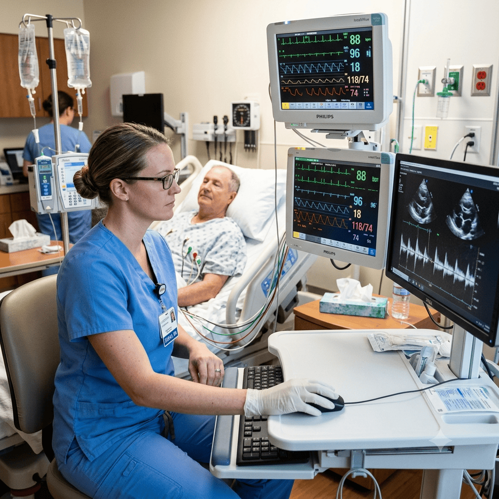
Diagnostic confirmation typically relies on echocardiography, cardiac MRI, or left ventriculography, which visualize the dyskinetic, bulging segment directly. Transthoracic echocardiography remains the first-line diagnostic tool because it is fast, noninvasive, and widely available at the bedside, allowing the healthcare team to assess wall motion, ejection fraction, and the presence of mural thrombus in a single study.
Medical Management Considerations
Alongside close monitoring, medical management plays a central role in limiting further ventricular remodeling. Providers commonly prescribe:
- ACE inhibitors or ARBs to reduce afterload and slow the remodeling process
- Beta-blockers to decrease myocardial oxygen demand and reduce dysrhythmia risk
- Anticoagulants, such as warfarin, when mural thrombus is identified or ejection fraction is significantly reduced
- Diuretics if concurrent heart failure symptoms are present
The nurse plays a key role in medication reconciliation, monitoring for therapeutic and adverse effects, and reinforcing why strict adherence to these regimens is essential for preventing further myocardial stress. Patients who understand the rationale behind their medications are far more likely to remain compliant after discharge, which directly reduces readmission rates for heart failure and dysrhythmia complications.
Nursing Interventions and Priority Care
Nursing management of a patient with a ventricular aneurysm after MI focuses on reducing cardiac workload, preventing complications, and monitoring for deterioration. Key interventions include:
- Continuous cardiac monitoring to detect life-threatening dysrhythmias early
- Strict blood pressure control, since elevated afterload increases wall stress on the weakened myocardium
- Activity pacing and rest periods to reduce myocardial oxygen demand during the healing phase
- Anticoagulation monitoring in patients started on therapy to prevent mural thrombus and embolic stroke
- Patient education on recognizing worsening heart failure symptoms and when to seek care
- Preparing patients for possible surgical aneurysmectomy if the aneurysm is large, symptomatic, or causing refractory dysrhythmias or heart failure
Every cardiac nursing bundle used on a telemetry or step-down unit should include standardized post-MI monitoring parameters, daily weight tracking, and clear criteria for escalating care — these bundles are critical tools for standardizing safe, evidence-based recovery care and reducing preventable complications.
Complications to Monitor
The three most dangerous complications of an untreated or unmonitored ventricular aneurysm are:
- Heart failure from loss of effective contractile mass
- Thromboembolism, including stroke, from stagnant blood pooling in the aneurysm sac
- Ventricular rupture, a rare but often fatal event, particularly with pseudoaneurysms
Prompt recognition and reporting of new symptoms by the bedside nurse is often the first line of defense against these outcomes.
💡 NCLEX Tips for Ventricular Aneurysm After MI
- Remember: persistent ST elevation after the acute MI phase strongly suggests aneurysm formation, not ongoing ischemia alone.
- Anterior wall MIs carry the highest risk for ventricular aneurysm — always correlate ECG leads V1-V4 with this complication.
- Prioritize dysrhythmia monitoring and blood pressure control as your top nursing actions.
- Mural thrombus risk means anticoagulation and neurological checks are essential safety priorities.
- Surgical aneurysmectomy is reserved for symptomatic, large, or complicated aneurysms — know this indication for exam questions.
Quick Reference Table
| Feature | True Ventricular Aneurysm | Pseudoaneurysm |
|---|---|---|
| Wall composition | All three myocardial layers (thinned/scarred) | Pericardium and thrombus only |
| Rupture risk | Low | High |
| Common location | Left ventricular apex/anterior wall | Inferior or posterior wall |
| Typical cause | Anterior MI, LAD occlusion | Contained free wall rupture |
| Management | Medical management or elective repair | Urgent surgical repair |
Conclusion
A ventricular aneurysm after MI represents a serious structural complication that demands vigilant nursing assessment, prompt recognition of warning signs, and coordinated care with the cardiology team. For every RN nurse working in cardiac care, mastering the pathophysiology, risk factors, and priority interventions for this condition builds both clinical confidence and exam readiness. Strengthen your understanding further by practicing related NCLEX questions and reviewing evidence-based cardiac nursing bundles to reinforce these critical concepts. Continue building your cardiac knowledge base with more practice questions at rn-nurse.com/nclex-qcm/ or explore in-depth training through our nursing courses.