
Introduction
Venous thromboembolism quietly threatens almost every hospitalized patient, and that’s exactly why it deserves more nursing attention than it often gets. Because up to half of DVT cases show no specific signs or symptoms at all, prevention and systematic risk assessment matter far more than waiting to catch obvious warning signs. This guide covers what actually drives VTE risk, how prophylaxis works, and what to do when recognition becomes an emergency.
Table of Contents
- What Is VTE? DVT and PE Explained
- Virchow’s Triad and Risk Factors
- Signs and Symptoms of DVT
- Signs and Symptoms of PE
- Nursing Assessment
- Diagnostic Workup
- VTE Prophylaxis: Mechanical and Pharmacologic
- Nursing Interventions for Suspected VTE
- Treatment
- Complications
- NCLEX Tips and Memory Tricks
- Clinical Pearls
- Key Takeaways
- FAQs
- References
What Is VTE? DVT and PE Explained
Venous thromboembolism (VTE) is an umbrella term covering two closely related conditions along the same disease spectrum. Deep vein thrombosis (DVT) is a blood clot forming in a deep vein, most often in the lower extremities. Pulmonary embolism (PE) occurs when part or all of that clot breaks free and travels to the lungs, where it can obstruct pulmonary blood flow. Because DVT and PE share the same underlying pathophysiology, hospitals generally approach prevention as a single combined VTE prevention strategy rather than treating the two conditions separately.
Virchow’s Triad and Risk Factors
Three underlying mechanisms, together known as Virchow’s triad, drive clot formation:
- Venous stasis — immobility, prolonged bed rest, long travel, paralysis
- Endothelial injury — surgery, trauma, central venous catheters
- Hypercoagulability — malignancy, pregnancy, certain medications (including hormonal therapy), inherited clotting disorders
Additional risk factors layer on top of this framework, including advanced age, obesity, prior VTE history, and major orthopedic or abdominal surgery — all of which is why individualized, structured risk assessment on admission genuinely matters, rather than applying a one-size-fits-all approach.
Signs and Symptoms of DVT
Because clinical signs alone are notoriously unreliable, up to half of patients with confirmed acute DVT lack specific symptoms entirely. When present, findings may include:
- Unilateral leg swelling
- Localized warmth and erythema
- Tenderness or pain along the affected vein
- A palpable cord in some cases
Since Homans sign (calf pain with dorsiflexion) is neither sensitive nor specific and can occur without any clot present at all, it’s no longer considered a reliable diagnostic finding on its own — it shouldn’t be relied on to rule DVT in or out.
Signs and Symptoms of PE
PE can range from clinically silent to immediately life-threatening. Watch for:
- Sudden-onset dyspnea
- Pleuritic chest pain
- Tachycardia
- Tachypnea
- Cough, sometimes with hemoptysis
- Anxiety or a sense of impending doom
- Hypoxia
- In massive PE: hypotension, syncope, or cardiac arrest
Any combination of these findings in an at-risk patient — especially sudden dyspnea with tachycardia — should prompt immediate evaluation rather than a wait-and-see approach.
Nursing Assessment
- Structured VTE risk assessment on admission and at defined intervals throughout the stay, since risk can change with a patient’s clinical course
- Bleeding risk assessment alongside VTE risk, since this determines whether pharmacologic prophylaxis is appropriate
- Regular skin and extremity checks for swelling, warmth, or tenderness
- Respiratory assessment, including oxygen saturation trends
- Mobility status and barriers to ambulation
Diagnostic Workup
- D-dimer, useful primarily for its high negative predictive value in low-risk patients
- Doppler ultrasound of the affected extremity for suspected DVT
- CT pulmonary angiogram, the primary imaging study for suspected PE
- Ventilation-perfusion (V/Q) scan, an alternative when CT angiogram isn’t appropriate (for example, in significant renal impairment or contrast allergy)
- ECG and chest X-ray, mainly to help rule out other causes of the presenting symptoms
VTE Prophylaxis: Mechanical and Pharmacologic
Because VTE prophylaxis prevents a genuinely dangerous and sometimes fatal complication, it’s treated as a required part of admission assessment in most hospitals, not an optional add-on. Prevention strategies include:
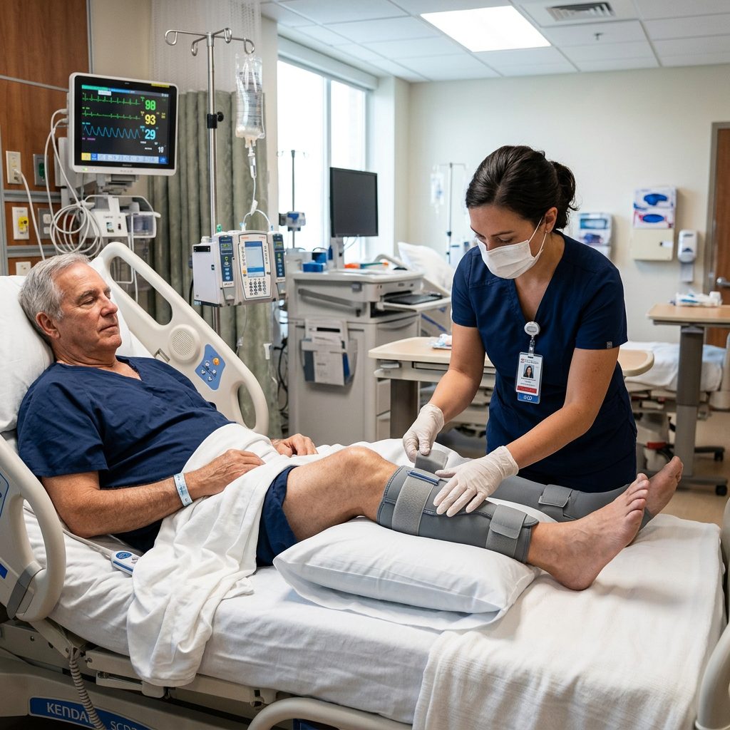
- Mechanical prophylaxis: sequential compression devices (SCDs) and graduated compression stockings, particularly important for patients with contraindications to anticoagulation
- Pharmacologic prophylaxis: subcutaneous unfractionated heparin or low molecular weight heparin, based on individualized risk-benefit assessment
- Early mobilization, which remains one of the simplest and most effective nursing-driven interventions available
- Patient and family education on the importance of prophylaxis, since patients sometimes resist SCDs or early ambulation without understanding why they matter
Notably, discharge-level compliance with extended prophylaxis (for example, after major cancer or orthopedic surgery) tends to be far weaker than inpatient compliance, which is a genuine gap nurses can help close through clear discharge teaching and follow-up coordination.
Nursing Interventions for Suspected VTE
- If PE is suspected, treat it as a medical emergency: notify the provider immediately, apply supplemental oxygen, and prepare for possible rapid response activation
- Keep the patient on strict bed rest if DVT is suspected, to reduce the risk of embolization, until cleared by the provider
- Never massage a suspected clot or affected extremity
- Continuously monitor vital signs and oxygen saturation in a patient with suspected PE
- Prepare for anticoagulation or thrombolytic therapy as ordered, and monitor closely for bleeding once started
Treatment
Once VTE is confirmed, treatment centers on anticoagulation — using heparin products, direct oral anticoagulants, or warfarin depending on the clinical picture — to prevent clot extension and further embolization. Thrombolytic therapy may be used in select cases of massive PE with hemodynamic instability. Inferior vena cava filters are generally reserved for patients who can’t tolerate anticoagulation, rather than used routinely.
Complications
- Pulmonary embolism progressing to right heart strain, hemodynamic collapse, or cardiac arrest
- Post-thrombotic syndrome (chronic leg swelling, pain, and skin changes following DVT)
- Chronic thromboembolic pulmonary hypertension
- Bleeding complications from anticoagulant therapy
- Recurrent VTE
NCLEX Tips and Memory Tricks
- Remember: up to half of DVT cases show no specific symptoms — don’t rely on obvious signs to justify skipping prophylaxis.
- Sudden dyspnea + tachycardia + pleuritic chest pain in an at-risk patient = think PE first.
- If PE is suspected, the priority nursing action is oxygen and provider notification, not waiting for confirmatory imaging.
- Mnemonic — “CLOT”: Check risk factors on admission, Limit immobility, Observe for DVT/PE signs, Treat suspected PE as an emergency.
Clinical Pearls
- Homans sign has fallen out of favor for good reason — a positive result doesn’t confirm DVT, and a negative result doesn’t rule it out.
- VTE prophylaxis compliance tends to drop sharply after discharge, so clear, specific discharge teaching (not just “keep taking your blood thinner”) genuinely closes a real care gap.
- A patient who “looks fine” on room air can still be developing a submassive PE — trend oxygen saturation and respiratory rate, not just a single reading.
Key Takeaways
- VTE encompasses DVT and PE, driven by venous stasis, endothelial injury, and hypercoagulability (Virchow’s triad).
- Up to half of DVT cases lack specific symptoms, making systematic risk assessment and prophylaxis essential.
- Prophylaxis combines mechanical measures (SCDs, early mobilization) with pharmacologic options (heparin products) based on individualized risk.
- Suspected PE is a medical emergency requiring immediate oxygen, provider notification, and close monitoring.
FAQs
What are the signs and symptoms of a pulmonary embolism?
Sudden dyspnea, pleuritic chest pain, tachycardia, tachypnea, cough (sometimes with hemoptysis), and anxiety or a sense of impending doom; massive PE can cause hypotension or cardiac arrest.
What is Virchow’s triad?
The three underlying mechanisms of clot formation: venous stasis, endothelial injury, and hypercoagulability.
What is the difference between mechanical and pharmacologic VTE prophylaxis?
Mechanical prophylaxis uses devices like sequential compression devices or compression stockings; pharmacologic prophylaxis uses anticoagulant medications like heparin products, chosen based on individualized bleeding risk.
What should a nurse do if a patient shows signs of a PE?
Notify the provider immediately, apply supplemental oxygen, monitor vital signs and oxygen saturation closely, and prepare for possible rapid response activation.
Is Homans sign a reliable way to detect DVT?
No. It’s neither sensitive nor specific and can occur without any clot present, so it shouldn’t be relied on to confirm or rule out DVT.
References
- NursingCE Central — VTE Prophylaxis Course
- AORN Journal — Guidelines in Practice: Prevention of Venous Thromboembolism
- Medscape — Venous Thromboembolism (VTE) Guidelines (2021 ACCP/CHEST, 2021 ESVS, 2020 ASH)
- Cureus — The Clot Stops Here: Insights Into Venous Thromboembolism Prophylaxis Adherence
RELATED ARTICLES:
- https://rn-nurse.com/atrial-fibrillation-nursing-rate-rhythm-control/
- https://rn-nurse.com/bariatric-surgery-nursing-considerations/
- https://rn-nurse.com/pacu-nursing-priorities-first-hour/
- https://rn-nurse.com/preoperative-nursing-assessment-checklist/
- https://rn-nurse.com/high-alert-medications-nurses/
- https://rn-nurse.com/lab-value-cheats/
- https://rn-nurse.com/fall-precautions-nursing/
- https://rn-nurse.com/safe-mobility-high-fall-risk-patients/
- https://rn-nurse.com/sepsis-management/
- https://rn-nurse.com/central-line-care/