Electrolytes keep your patient’s heart beating in a healthy rhythm. When potassium, calcium, or magnesium levels get too high or too low, the EKG shows early warning signs before a patient’s heart goes into a dangerous rhythm.
If you’re a nurse, you must know how these changes look on the EKG strip. Let’s break this down step-by-step so you’ll feel confident spotting trouble early.
📌 Why Electrolytes Matter for the Heart
Electrolytes are charged particles in the blood that help electrical signals move through the heart muscle. They control how the heart contracts and resets for the next beat.
The Big 3 to watch:
✅ Potassium (K+) — major player in cardiac electrical conduction.
✅ Calcium (Ca++) — affects contraction strength and repolarization.
✅ Magnesium (Mg++) — stabilizes heart cells and works with potassium.
When any of these get out of balance, the electrical signals get messy. This can cause arrhythmias — irregular, fast, or slow heart rhythms — and can lead to cardiac arrest.
🔑 Quick Normal Lab Ranges
| Electrolyte | Normal Range |
|---|---|
| Potassium | 3.5–5.0 mEq/L |
| Calcium | 8.5–10.5 mg/dL (total), 4.5–5.5 mg/dL (ionized) |
| Magnesium | 1.5–2.5 mEq/L |
Keep these in mind while reading EKG strips!
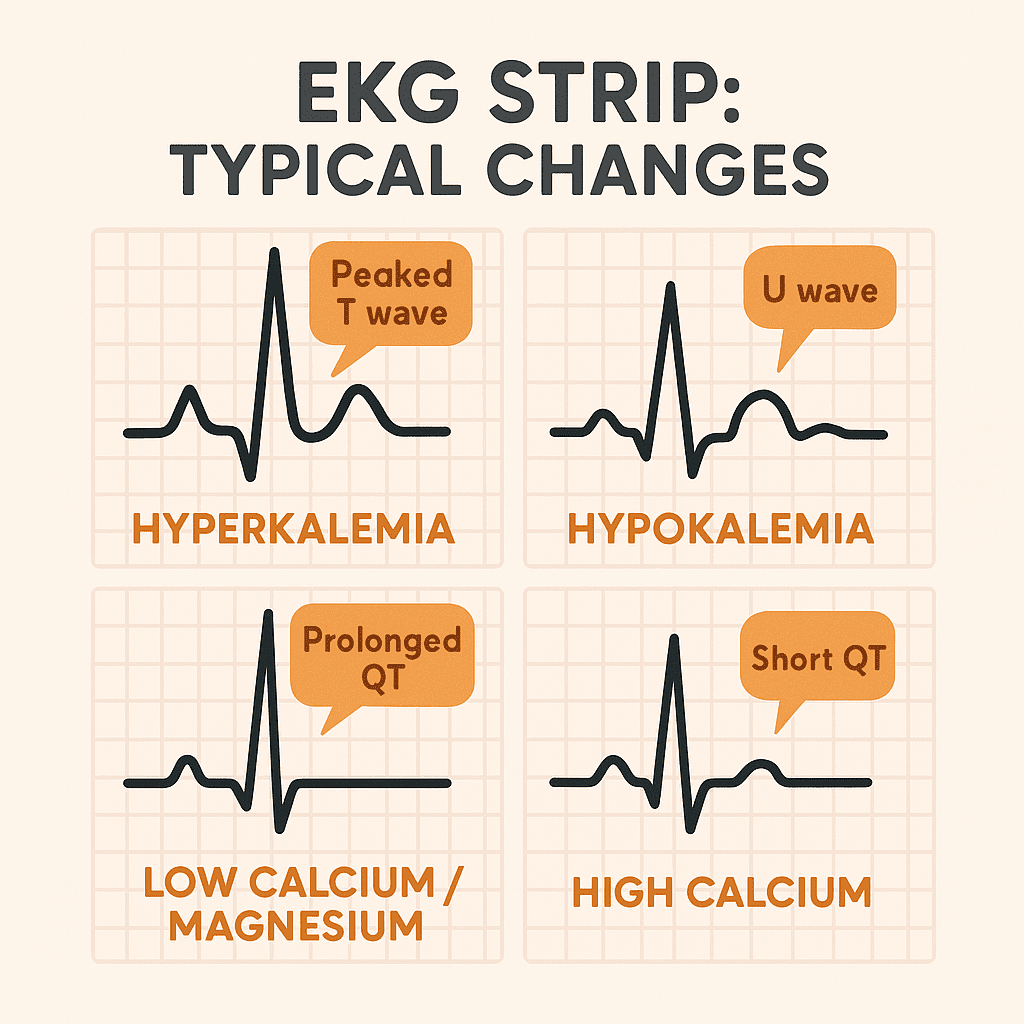
1️⃣ Potassium Imbalances and the EKG
Potassium is the biggest player for cardiac rhythms. Too much or too little can be deadly.
✅ Hypokalemia (Low Potassium)
Causes:
- Diuretics (like furosemide)
- Vomiting or diarrhea
- Poor intake
EKG Changes:
- Flat or inverted T waves
- U waves appear (extra wave after T wave)
- ST depression
- Possible PVCs (premature ventricular contractions)
Why it’s dangerous:
Low potassium makes the heart irritable and prone to dangerous arrhythmias like VTach or VFib.
Nursing actions:
- Check potassium levels.
- Replace potassium slowly — never IV push!
- Monitor EKG continuously.
- Treat underlying cause (stop wasting diuretics if possible).
✅ Hyperkalemia (High Potassium)
Causes:
- Kidney failure
- Tissue damage (burns, trauma)
- Certain meds (potassium-sparing diuretics, ACE inhibitors)
EKG Changes:
- Tall, peaked T waves (early sign!)
- Widened QRS complex
- Prolonged PR interval
- Flattened or absent P wave
- Can lead to sine wave pattern and asystole
Why it’s dangerous:
High potassium slows conduction, leading to heart block, cardiac arrest, or asystole.
Nursing actions:
- Verify lab result — hemolyzed sample can show false high.
- Give calcium gluconate (stabilizes heart).
- Give insulin and glucose (pushes K+ into cells).
- Consider diuretics or dialysis if severe.
- Monitor EKG closely.
2️⃣ Calcium Imbalances and the EKG
Calcium affects the plateau phase of the heart’s electrical cycle. It impacts the QT interval most.
✅ Hypocalcemia (Low Calcium)
Causes:
- Low parathyroid hormone
- Vitamin D deficiency
- Kidney disease
EKG Changes:
- Prolonged QT interval
- Possible Torsades de Pointes (deadly!)
Why it’s dangerous:
Long QT means the ventricles take too long to repolarize — increasing risk for sudden arrhythmias.
Nursing actions:
- Give calcium gluconate or calcium chloride IV.
- Monitor for muscle cramps or tetany.
- Watch for signs of Torsades (sudden rapid rhythm).
✅ Hypercalcemia (High Calcium)
Causes:
- Hyperparathyroidism
- Bone cancer
- Excess vitamin D
EKG Changes:
- Shortened QT interval
- Possible widened T waves
Why it’s dangerous:
Too much calcium speeds up repolarization, which can cause bradycardia or heart block in severe cases.
Nursing actions:
- Hydrate the patient.
- Give IV fluids and loop diuretics to flush calcium.
- Treat the cause (e.g., parathyroid issue).
3️⃣ Magnesium Imbalances and the EKG
Magnesium works behind the scenes to keep potassium balanced and stabilize cell membranes.
✅ Hypomagnesemia (Low Magnesium)
Causes:
- Alcoholism
- Malnutrition
- Diuretics
EKG Changes:
- Prolonged QT interval
- Possible Torsades de Pointes
- PVCs or VTach
Why it’s dangerous:
Low magnesium worsens low potassium effects — together, they’re a recipe for deadly arrhythmias.
Nursing actions:
- Replace magnesium IV slowly.
- Check potassium too — they often drop together.
- Watch for Torsades — treat immediately with IV magnesium sulfate.
✅ Hypermagnesemia (High Magnesium)
Causes:
- Kidney failure
- Excess antacid or laxative use (milk of magnesia)
EKG Changes:
- Prolonged PR interval
- Widened QRS
- Bradycardia
Why it’s dangerous:
High magnesium slows the heart and can lead to cardiac arrest in extreme cases.
Nursing actions:
- Stop magnesium intake.
- Give calcium gluconate to reverse severe effects.
- Dialysis if kidneys can’t clear it.
🗂️ Quick EKG Cheat Sheet
| Electrolyte | Low Level | High Level |
|---|---|---|
| Potassium | Flat T, U wave, ST depression | Peaked T, wide QRS, sine wave |
| Calcium | Prolonged QT | Short QT |
| Magnesium | Prolonged QT, Torsades | Prolonged PR, wide QRS, bradycardia |
✅ How Nurses Use This in Real Life
👉 Always check lab trends — don’t rely on a single number.
👉 Connect the dots: weird EKG + weird labs = take action fast!
👉 Know the meds that affect electrolytes — diuretics, ACE inhibitors, chemotherapy, supplements.
👉 Always treat the root cause — giving electrolytes is only part of the fix.
👉 In emergencies, keep calcium gluconate and magnesium sulfate ready — they save lives!