When caring for patients with heart conditions, understanding atrial rhythms is a must for every nurse. This guide will break down Atrial Fibrillation (AFib), Atrial Flutter (AFlutter), and Supraventricular Tachycardia (SVT) — how to spot them on the monitor, what causes them, what signs to watch for, and what actions to take.
📌 What Are Atrial Rhythms?
Atrial rhythms are heart rhythms that start in the atria — the upper chambers of the heart. When something disrupts the normal electrical path in the atria, it can cause the heart to beat too fast, too slow, or irregularly.
The three main atrial rhythms nurses must know are:
- Atrial Fibrillation (AFib)
- Atrial Flutter (AFlutter)
- Supraventricular Tachycardia (SVT)
❤️ 1. Atrial Fibrillation (AFib)
🔍 What Is It?
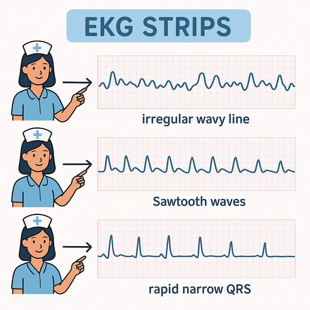
AFib is the most common sustained arrhythmia. In AFib, the atria quiver instead of beating in a coordinated way. This causes an irregular and often fast heart rate.
🩺 How to Recognize AFib
On an EKG:
- No distinct P waves.
- Irregularly irregular R-R intervals.
- Wavy baseline (fibrillatory waves).
Patients may feel:
- Palpitations
- Shortness of breath
- Fatigue
- Dizziness
⚡ Causes of AFib
- High blood pressure
- Heart disease
- Recent heart surgery
- Hyperthyroidism
- Alcohol (holiday heart)
- Advanced age
🏥 Nursing Interventions for AFib
- Assess vital signs: Check for hypotension and decreased cardiac output.
- Monitor for clots: AFib increases stroke risk because blood pools in the atria.
- Administer meds: Beta-blockers, calcium channel blockers, or anticoagulants as ordered.
- Prepare for procedures: Patients may need cardioversion or ablation.
💙 2. Atrial Flutter (AFlutter)
🔍 What Is It?
AFlutter is similar to AFib but more organized. The atria beat very fast in a regular pattern — like a “sawtooth” flutter.
🩺 How to Recognize AFlutter
On an EKG:
- Sawtooth-shaped flutter waves (F waves).
- Regular or irregular ventricular response.
- Atrial rate usually 250–350 bpm.
Patients may report:
- Rapid heartbeat
- Weakness
- Lightheadedness
⚡ Causes of AFlutter
- Heart surgery
- Heart failure
- Valve disease
- Chronic lung disease
🏥 Nursing Interventions for AFlutter
- Monitor rate and rhythm: Watch for rapid ventricular rates.
- Administer medications: Similar to AFib — rate control and anticoagulation.
- Prepare for treatment: Cardioversion is often more successful in AFlutter than AFib.
- Educate the patient: Explain possible procedures like ablation.
💚 3. Supraventricular Tachycardia (SVT)
🔍 What Is It?
SVT is a rapid heart rhythm that starts above the ventricles. It is often sudden and can stop suddenly too. It includes AVNRT and AVRT.
🩺 How to Recognize SVT
On an EKG:
- Rate: 150–250 bpm.
- P waves hidden in the T waves.
- Narrow QRS.
Patients may feel:
- Sudden racing heart
- Anxiety
- Chest tightness
- Shortness of breath
⚡ Causes of SVT
- Stimulants (caffeine, drugs)
- Stress
- Electrolyte imbalance
- Overexertion
🏥 Nursing Interventions for SVT
- Stay calm and reassure the patient.
- Try vagal maneuvers: Instruct patient to bear down or cough.
- Prepare for medication: Adenosine is often given to break the rhythm.
- Monitor continuously: Watch for recurrence.
- Prepare for synchronized cardioversion if patient is unstable.
🗂️ Key Differences
| Rhythm | EKG Feature | Atrial Rate | Ventricular Response | Common Treatment |
|---|---|---|---|---|
| AFib | Irregular, no P waves | 350–600 | Irregularly irregular | Rate control, anticoagulants |
| AFlutter | Sawtooth F waves | 250–350 | Regular or irregular | Cardioversion, rate control |
| SVT | Rapid, narrow QRS | N/A | Regular | Vagal maneuvers, adenosine |
⚕️ Why Nurses Must Master Atrial Rhythms
Understanding atrial rhythms helps nurses:
- Spot complications early.
- Prevent strokes by ensuring patients receive anticoagulation.
- Respond fast in emergencies.
- Educate patients on signs and triggers.
💡 Quick Tips for Nurses
✅ Know normal EKG strips — it makes abnormal strips stand out.
✅ Always check the patient, not just the monitor.
✅ Learn and practice rhythm interpretation regularly.
✅ Communicate clearly with your team about any changes.
📝 Conclusion
AFib, AFlutter, and SVT are common but manageable when recognized early. By understanding how they appear on EKGs and what signs to watch for, you can protect your patient’s heart and brain.
Stay curious, keep practicing your EKG reading skills, and you’ll feel more confident when these rhythms show up in your patients!