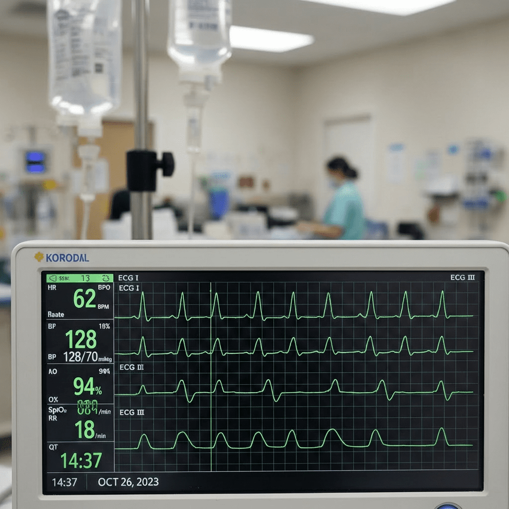
Electrolyte imbalances can silently change the ECG before symptoms become severe. For any nurse or registered nurse, recognizing these early cardiac clues can prevent cardiac arrest.
Advanced ECG interpretation in electrolyte emergencies is heavily tested on the NCLEX and frequently seen in critical care and telemetry nursing practice.
When electrolytes shift, the heart’s electrical system is often the first to show danger signs.
Whether you are reviewing your cardiac chapter inside a comprehensive nursing bundle or preparing for your next shift as an RN nurse, this guide simplifies advanced ECG patterns linked to electrolyte disorders.
Why ECG Changes Matter in Electrolyte Emergencies
Electrolytes regulate:
- Cardiac depolarization
- Repolarization
- Conduction velocity
- Myocardial contractility
Even small abnormalities in potassium, calcium, or magnesium can cause life-threatening dysrhythmias.
For the registered nurse, early ECG recognition = early intervention.
1️⃣ Hyperkalemia: The Most Dangerous ECG Pattern
Hyperkalemia is one of the most critical electrolyte emergencies tested on the NCLEX.
Key ECG Changes (Progressive Pattern)
Early:
- Tall, peaked T waves
- Shortened QT interval
Moderate:
- Flattened or absent P waves
- Prolonged PR interval
- Widened QRS
Severe:
- Sine-wave pattern
- Ventricular fibrillation
- Asystole
Advanced Interpretation Tip
Hyperkalemia affects the ECG globally, not regionally. This helps the RN nurse differentiate it from myocardial infarction.
2️⃣ Hypokalemia: Irritable Myocardium
Hypokalemia increases the risk of dangerous arrhythmias, especially in cardiac patients.
Classic ECG Findings
- Flattened T waves
- Prominent U waves
- ST depression
- Prolonged QT interval
Why It Matters in Nursing Practice
Hypokalemia can trigger:
- Atrial fibrillation
- Ventricular tachycardia
- Torsades de pointes
On the NCLEX, hypokalemia questions often involve patients on diuretics.
3️⃣ Hypercalcemia: Short QT Interval
Calcium directly affects myocardial contraction.
ECG Changes
- Shortened QT interval
- Possible bradycardia
Clinical Clues
- Kidney stones
- Confusion
- Bone pain
For a registered nurse, recognizing a short QT interval can point toward hypercalcemia quickly.
4️⃣ Hypocalcemia: Prolonged QT Interval
Hypocalcemia increases cardiac excitability.
ECG Findings
- Prolonged QT interval
- Risk for torsades de pointes
Nursing Priority
Monitor telemetry closely. Seizure precautions may also be necessary.
This is a high-yield concept in most comprehensive nursing bundle cardiac sections.
5️⃣ Magnesium Imbalances
Magnesium abnormalities are frequently overlooked but dangerous.
Hypomagnesemia
- Prolonged QT
- Torsades de pointes
- Ventricular arrhythmias
Hypermagnesemia
- Bradycardia
- Prolonged PR interval
- Widened QRS (severe cases)
Magnesium is often linked to potassium abnormalities, so experienced RN nurses always assess both.
Advanced ECG Comparison Table for NCLEX Review
| Electrolyte | Key ECG Change | Dangerous Rhythm Risk |
|---|---|---|
| Hyperkalemia | Peaked T waves, wide QRS | VF, asystole |
| Hypokalemia | U waves, flat T | Torsades, VT |
| Hypercalcemia | Short QT | Bradycardia |
| Hypocalcemia | Long QT | Torsades |
| Hypomagnesemia | Long QT | Torsades |
This table is ideal for quick revision before your NCLEX exam or clinical shift.
How Registered Nurses Should Approach ECG Changes
Advanced ECG interpretation requires a systematic method:
Step 1: Assess the Rate and Rhythm
Is it sinus? Is there bradycardia or tachycardia?
Step 2: Evaluate Intervals
- PR interval
- QRS width
- QT interval
Step 3: Examine T Waves Carefully
Electrolyte emergencies frequently alter T-wave morphology.
Step 4: Correlate With Labs
Never interpret ECG findings in isolation.
A skilled nurse always combines ECG data with potassium, calcium, and magnesium lab values.
NCLEX-Style Practice Question
A patient receiving loop diuretics develops muscle weakness. ECG shows flattened T waves and prominent U waves. What should the nurse suspect?
A. Hyperkalemia
B. Hypokalemia
C. Hypercalcemia
D. Hypocalcemia
Correct Answer: B
This pattern is classic for hypokalemia and commonly tested in nursing exams.
Emergency Nursing Priorities in Electrolyte Imbalances
For every registered nurse:
- Place patient on cardiac monitor
- Obtain stat electrolyte labs
- Prepare emergency medications
- Notify provider immediately
- Follow ACLS protocols if unstable
Rapid recognition is essential in critical care nursing.
Final Takeaways for Nurses and RN Nurses
Advanced ECG interpretation in electrolyte emergencies is a powerful clinical skill.
Remember:
- Potassium changes = T wave changes
- Calcium changes = QT interval changes
- Magnesium changes = arrhythmia risk
- Always assess globally vs regionally
Mastering these ECG patterns improves your confidence as an RN nurse, strengthens clinical decision-making, and increases your success on the NCLEX.
If you’re building your knowledge through a structured nursing bundle, make sure electrolyte ECG interpretation is a core focus area.