
Introduction
Diabetic foot ulcers (DFUs) are one of the most common and costly complications of diabetes mellitus, and they are a leading cause of non-traumatic lower-limb amputation worldwide. For nursing students and practicing nurses alike, understanding how to assess, stage, and manage these wounds — and how to teach patients to prevent them in the first place — is essential clinical knowledge tested heavily on the NCLEX and used daily on medical-surgical, endocrine, and wound care units.
This guide walks through the pathophysiology behind diabetic foot ulcers, how to perform a thorough nursing assessment, the staging systems used to classify severity, evidence-based interventions, and the patient education strategies that actually reduce ulcer recurrence and amputation risk.
Table of Contents
- Definition and Overview
- Causes and Risk Factors
- Pathophysiology
- Signs and Symptoms
- Nursing Assessment
- Diagnosis and Staging (Wagner Classification)
- Laboratory Tests and Diagnostic Imaging
- Medical and Surgical Treatment
- Pharmacology
- Nursing Interventions
- Patient Education
- Complications
- Prevention
- Prognosis
- NCLEX Tips
- Memory Tricks
- Clinical Pearls
- Key Takeaways
- FAQs
- References
Definition
A diabetic foot ulcer is a full-thickness break in the skin, most often on the plantar (bottom) surface of the foot, occurring in a person with diabetes mellitus. It develops from a combination of nerve damage, poor circulation, and repeated unnoticed pressure or trauma. Left untreated, DFUs can progress to deep tissue infection, osteomyelitis, and lower-extremity amputation.
Causes and Risk Factors
DFUs rarely have a single cause — they result from the interaction of several diabetes-related complications:
- Peripheral neuropathy — loss of protective sensation means patients often don’t feel cuts, blisters, or pressure points forming.
- Peripheral arterial disease (PAD) — reduced blood flow slows healing and increases tissue breakdown risk.
- Poor glycemic control — chronic hyperglycemia impairs white blood cell function, collagen synthesis, and microvascular circulation.
- Foot deformities — bunions, hammertoes, and Charcot foot create abnormal pressure points.
- Improper footwear — ill-fitting shoes are a frequent trigger for callus and ulcer formation.
- Prior history of ulceration or amputation — the single strongest predictor of a new ulcer.
- Long duration of diabetes — risk increases the longer a patient has lived with the disease.
- Smoking — worsens peripheral circulation and impairs healing.
- Visual impairment or limited mobility — reduces a patient’s ability to inspect their own feet.
- Renal disease (diabetic nephropathy) — associated with poorer wound healing outcomes.
Pathophysiology
Chronic hyperglycemia damages both peripheral nerves and small blood vessels over time. Sensory neuropathy causes loss of pain perception, so minor trauma — a pebble in a shoe, an ill-fitting sock seam, a hot bath — goes unnoticed and unaddressed. Motor neuropathy weakens the small intrinsic muscles of the foot, leading to structural deformities that create new areas of high pressure, particularly over the metatarsal heads. Autonomic neuropathy reduces sweating, leaving skin dry, cracked, and more prone to fissures that serve as entry points for bacteria.
At the same time, peripheral arterial disease reduces blood flow to the extremities, so any break in the skin heals slowly. Chronic hyperglycemia also impairs neutrophil function and collagen deposition, further delaying wound closure. This combination — insensate skin, abnormal pressure, poor circulation, and impaired healing — sets the stage for a minor injury to progress into a chronic, non-healing ulcer.
Signs and Symptoms
- Open wound or break in skin, often on the plantar surface, heel, or over bony prominences
- Callus formation surrounding the wound
- Drainage or exudate (may be absent early on)
- Odor, if infection is present
- Redness, warmth, or swelling around the wound edges
- Black or necrotic tissue at the wound base in advanced cases
- Notably, many patients report little to no pain due to underlying neuropathy — this absence of pain should never be mistaken for absence of severity
Nursing Assessment
A comprehensive diabetic foot assessment should be performed at every diabetes-related visit and upon any hospital admission for a patient with diabetes. Key components include:
Visual inspection
- Inspect between the toes, the plantar surface, heels, and around nail beds
- Note skin color, temperature, hair distribution, and nail condition
- Check for calluses, fissures, blistering, or existing ulceration
Neurological assessment
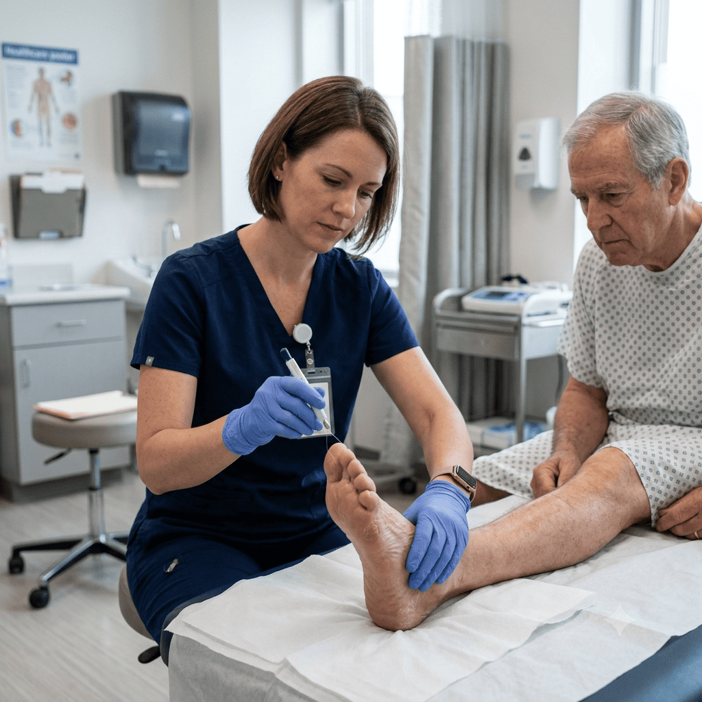
- Perform the 10-gram monofilament test at multiple plantar sites to assess protective sensation
- Assess vibration sense with a 128-Hz tuning fork
- Test deep tendon reflexes (ankle jerk is often diminished first)
Vascular assessment
- Palpate dorsalis pedis and posterior tibial pulses bilaterally
- Assess capillary refill (should be under 3 seconds)
- Note skin temperature and any dependent rubor or pallor on elevation
- An ankle-brachial index (ABI) may be ordered if PAD is suspected
Wound assessment (if an ulcer is present)
- Measure length, width, and depth
- Assess wound bed tissue: granulation, slough, or eschar
- Note the amount, color, and odor of exudate
- Assess the periwound skin for maceration, erythema, or induration
- Classify the ulcer using a standardized staging tool (see below)
Footwear and gait assessment
- Inspect the patient’s shoes for areas of excessive wear
- Observe gait for abnormal weight distribution
Diagnosis and Staging: Wagner Classification
The Wagner Ulcer Classification System is the most widely used staging tool in nursing and clinical practice:
| Grade | Description |
|---|---|
| 0 | Intact skin; foot at risk (no open lesion) |
| 1 | Superficial ulcer involving only the epidermis/dermis |
| 2 | Deeper ulcer extending into tendon, ligament, joint capsule, or fascia |
| 3 | Deep ulcer with abscess, osteomyelitis, or joint sepsis |
| 4 | Localized gangrene (e.g., toe, forefoot) |
| 5 | Extensive gangrene involving the entire foot |
Other tools nurses may encounter include the University of Texas Classification System, which adds infection and ischemia grading to depth, and the PEDIS system used more often in research settings.
Laboratory Tests and Diagnostic Imaging
- HbA1c — reflects long-term glycemic control
- Fasting blood glucose / point-of-care glucose — monitors current control
- CBC with differential — elevated WBCs may indicate infection
- ESR and CRP — inflammatory markers, useful in monitoring infection or suspected osteomyelitis
- Wound culture — obtained via tissue biopsy or curettage (not superficial swab, which often reflects surface colonization rather than true infection)
- X-ray of the foot — screens for bone involvement or gas in soft tissue
- MRI — gold standard for detecting osteomyelitis
- Ankle-brachial index (ABI) — evaluates arterial perfusion
Medical Treatment
Treatment is guided by wound grade, presence of infection, and vascular status:
- Debridement — removal of necrotic or non-viable tissue (sharp, enzymatic, mechanical, or autolytic) to promote a healthy wound bed
- Infection control — culture-guided antibiotic therapy for confirmed infection
- Offloading — total contact casting, removable cast walkers, or specialized footwear to redistribute pressure away from the ulcer
- Revascularization — angioplasty or bypass surgery when significant PAD is contributing to poor healing
- Advanced wound therapies — negative pressure wound therapy (NPWT), bioengineered skin substitutes, or hyperbaric oxygen therapy for select non-healing wounds
- Surgical management — may include incision and drainage of abscesses or, in severe cases, partial or full amputation
Pharmacology
- Antibiotics — selected based on culture and sensitivity; commonly used agents include those covering both gram-positive and gram-negative organisms in moderate-to-severe infections
- Insulin or oral hypoglycemics — tight glycemic control supports wound healing
- Topical antimicrobial dressings — silver-impregnated or iodine-based dressings for wounds with high bioburden
- Analgesics — used judiciously, since many patients have reduced pain sensation; pain that does emerge in a previously insensate wound can be a red flag for worsening infection
Nursing Interventions
- Perform and document a thorough foot and wound assessment on every shift for hospitalized patients
- Maintain a moist wound healing environment with appropriate dressing selection based on exudate level
- Reinforce and assist with offloading devices; monitor adherence
- Monitor blood glucose levels closely and coordinate with the care team on insulin or medication adjustments
- Administer prescribed antibiotics and monitor for therapeutic response
- Assess for signs of worsening infection: increasing erythema, warmth, purulent drainage, fever, or new-onset pain in a previously insensate area
- Encourage smoking cessation and provide resources
- Collaborate with podiatry, vascular surgery, wound care specialists, and diabetes educators
- Ensure adequate nutrition, including protein intake, to support tissue repair
- Position and reposition to relieve pressure on the affected limb
Patient Education
- Inspect both feet daily, including the soles and between toes — a mirror can help with hard-to-see areas
- Never walk barefoot, even indoors
- Wash feet daily with lukewarm (not hot) water and dry thoroughly, especially between the toes
- Moisturize the feet but avoid applying lotion between the toes
- Trim toenails straight across; avoid cutting into the corners
- Wear well-fitting, cushioned shoes and clean, seamless socks; check the inside of shoes for debris before putting them on
- Never attempt to self-treat calluses, corns, or ingrown nails with sharp instruments
- Report any redness, swelling, drainage, or new sore to a healthcare provider immediately, even if it doesn’t hurt
- Attend regular podiatry and diabetes follow-up appointments
- Maintain blood glucose targets as closely as possible
- Avoid smoking, which worsens circulation
Complications
- Wound infection and cellulitis
- Osteomyelitis (bone infection)
- Abscess formation
- Sepsis
- Gangrene
- Lower-extremity amputation
- Delayed healing and ulcer recurrence
- Reduced mobility and quality of life
Prevention
Prevention hinges on early identification of the at-risk foot and consistent daily self-care. Annual comprehensive foot exams, routine monofilament testing, patient education, appropriate footwear, and tight glycemic control are the pillars of DFU prevention. Multidisciplinary foot care programs involving nursing, podiatry, and diabetes education have been shown to significantly reduce ulcer incidence and amputation rates.
Prognosis
Prognosis depends heavily on ulcer severity at diagnosis, vascular status, glycemic control, and how quickly treatment begins. Superficial ulcers (Wagner Grade 1) often heal well with offloading and local wound care. Deeper ulcers involving bone or significant ischemia carry a substantially higher risk of amputation. Even after healing, DFUs have a high recurrence rate, making ongoing preventive foot care essential for life.
NCLEX Tips
- Remember that pain is often absent in diabetic foot ulcers due to neuropathy — don’t rule out a serious wound just because the patient reports minimal discomfort
- Know the Wagner Classification grades — NCLEX may present a wound description and ask you to identify the correct grade or appropriate intervention
- Offloading is a priority intervention — a question may test whether you recognize that continued weight-bearing on an ulcer will prevent healing regardless of dressing choice
- Distinguish between neuropathic ulcers (typically on plantar surfaces, painless, surrounded by callus) and ischemic/arterial ulcers (often on toes or lateral foot, painful, with poor pulses)
- Glycemic control questions often pair with wound-healing rationale — hyperglycemia impairs neutrophil function and delays healing
Memory Tricks
“FEET” for prevention teaching:
- F – Footwear check daily
- E – Examine feet daily (use a mirror)
- E – Every year, comprehensive podiatry exam
- T – Temperature of water should be lukewarm, never hot
Clinical Pearls
- A wound swab culture from the surface of a DFU frequently reflects colonizing bacteria, not the true infecting organism — deep tissue culture or bone biopsy is more reliable when osteomyelitis is suspected
- New-onset pain in a previously painless, neuropathic ulcer can signal worsening infection and warrants prompt reassessment
- Never debride a wound with significant arterial insufficiency without vascular workup first — restoring blood flow may need to precede aggressive debridement
- A callus surrounding an ulcer is a sign of ongoing repetitive pressure and should prompt reassessment of offloading strategy
Key Takeaways
- Diabetic foot ulcers result from the combined effects of neuropathy, peripheral arterial disease, and repetitive trauma
- The Wagner Classification is the standard staging tool nurses use to grade ulcer severity
- Offloading pressure is central to healing — no dressing can substitute for pressure relief
- Absence of pain does not mean absence of severity
- Daily foot inspection and patient education are the most effective prevention strategies
- Multidisciplinary care (nursing, podiatry, vascular surgery, endocrinology) improves outcomes and reduces amputation risk
FAQs
What is the best way to treat a diabetic foot ulcer? Treatment combines debridement of non-viable tissue, infection control, offloading of pressure from the wound, glycemic management, and — when needed — revascularization. The specific approach depends on the ulcer’s Wagner grade and whether infection or ischemia is present.
How can nurses prevent diabetic foot ulcers? Through routine, structured foot assessments (including monofilament testing), patient education on daily foot inspection and proper footwear, glycemic control support, and timely referral to podiatry for at-risk patients.
What are the stages of a diabetic foot ulcer? The Wagner Classification grades ulcers from 0 (intact but at-risk skin) to 5 (extensive gangrene), based on depth and the presence of infection or tissue death.
How do you assess a diabetic foot ulcer? Assessment includes visual inspection, neurological testing (monofilament and vibration sense), vascular assessment (pulses, capillary refill, ABI), and wound-specific measurements of size, depth, exudate, and periwound skin condition.
What does an infected diabetic ulcer look like? Signs include increased redness or warmth around the wound, purulent or foul-smelling drainage, swelling, new or worsening pain, and systemic signs such as fever in more severe cases.
Why do diabetics get foot ulcers? A combination of peripheral neuropathy (loss of protective sensation), peripheral arterial disease (reduced blood flow), and chronic hyperglycemia (impaired healing) makes the diabetic foot vulnerable to unnoticed injury that progresses into a chronic wound.
References
- Centers for Disease Control and Prevention (CDC) — Diabetes and Foot Complications
- National Institute of Diabetes and Digestive and Kidney Diseases (NIDDK)
- American Diabetes Association (ADA) — Standards of Care in Diabetes
- National Council of State Boards of Nursing (NCSBN) — NCLEX Test Plan
- Wound, Ostomy and Continence Nurses Society (WOCN)
- International Working Group on the Diabetic Foot (IWGDF) Guidelines