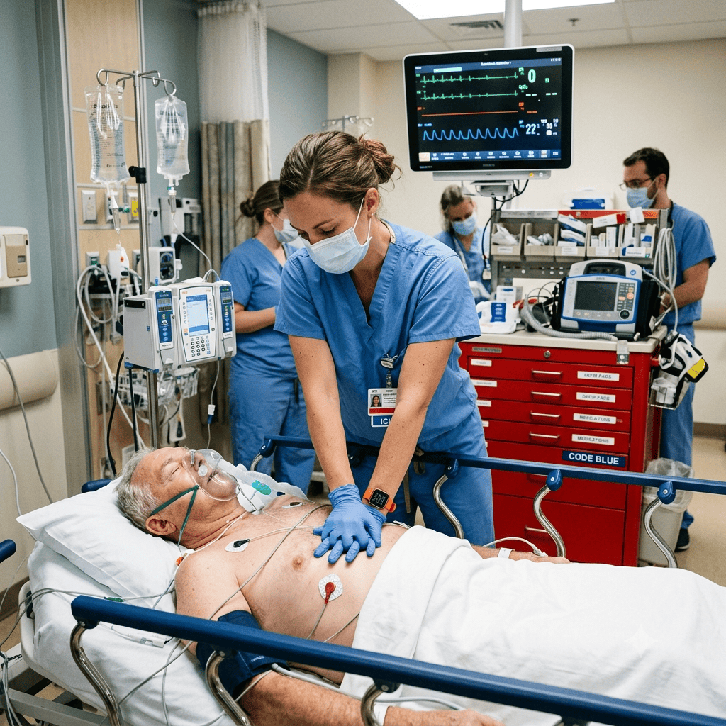
A patient’s monitor shows a perfectly organized rhythm, yet there is no pulse. This unsettling scenario is the hallmark of pulseless electrical activity (PEA), a cardiac arrest rhythm that demands immediate recognition and a systematic response. For any nurse working in critical care, the emergency department, or a med-surg floor, understanding pulseless electrical activity nursing management is not optional — it is a core competency tested heavily on the NCLEX and required in real-world code situations. Every registered nurse must be able to identify PEA, initiate high-quality CPR, and work through its reversible causes without hesitation.
PEA accounts for a significant share of in-hospital cardiac arrests, and unlike ventricular fibrillation, it is not a “shockable” rhythm. This means the nurse’s clinical reasoning — not a defibrillator — is often what saves the patient’s life.
What Is Pulseless Electrical Activity?
Pulseless electrical activity occurs when the heart’s electrical system generates an organized rhythm on the monitor, but the heart muscle fails to produce an effective mechanical contraction. In other words, there is electrical activity without a palpable pulse or measurable blood pressure. This is fundamentally different from asystole, where there is no electrical activity at all, and from ventricular fibrillation, where the rhythm is chaotic and shockable.
Key characteristics of PEA include:
- Organized rhythm visible on the cardiac monitor (may look like normal sinus, bradycardia, or a wide-complex rhythm)
- Absent pulse on palpation despite the rhythm
- No cardiac output detectable by Doppler or arterial line waveform
- Patient is unresponsive and apneic or agonal breathing
Because the rhythm can look deceptively normal, the RN nurse must always confirm a pulse before trusting the monitor alone — a critical NCLEX teaching point.
Common Causes: The H’s and T’s
Successful pulseless electrical activity nursing intervention hinges on rapidly identifying and correcting the underlying, reversible cause. The American Heart Association organizes these causes into the well-known H’s and T’s, a framework every nursing student should memorize for both clinical practice and the NCLEX.
The Five H’s:
- Hypovolemia — often the most common cause; treat with rapid IV fluid resuscitation
- Hypoxia — ensure airway patency and provide 100% oxygen
- Hydrogen ion (acidosis) — consider sodium bicarbonate per ACLS protocol
- Hyperkalemia/Hypokalemia — correct with calcium chloride, insulin/dextrose, or potassium replacement
- Hypothermia — initiate active rewarming measures
The Five T’s:
- Tension pneumothorax — needle decompression is life-saving
- Tamponade (cardiac) — pericardiocentesis relieves pressure on the heart
- Toxins — administer antidotes when a specific overdose is identified
- Thrombosis (pulmonary) — consider fibrinolytics for suspected massive PE
- Thrombosis (coronary) — treat as an acute MI once ROSC is achieved
Priority Nursing Actions During PEA
When a patient develops PEA, the nursing team must move through ACLS priorities without delay. Bold clinical judgment and clear role delegation are essential in this high-acuity scenario.
- Confirm unresponsiveness and absence of pulse — check for no more than 10 seconds
- Call for help and begin high-quality CPR immediately — compressions at a rate of 100–120/min, depth of at least 2 inches
- Secure the airway and provide ventilation with a bag-valve mask or advanced airway
- Establish IV/IO access for medication administration
- Administer epinephrine 1 mg IV/IO every 3–5 minutes per ACLS protocol
- Search for and treat the underlying H’s and T’s while compressions continue
- Reassess rhythm every 2 minutes, minimizing interruptions in compressions
A well-coordinated code team, guided by clear SBAR communication, dramatically improves the odds of achieving return of spontaneous circulation (ROSC).
Differentiating True PEA from Pseudo-PEA
Advanced practice increasingly distinguishes between true PEA, where there is no meaningful cardiac contraction at all, and pseudo-PEA, where the heart is still contracting weakly but too poorly to generate a palpable pulse or measurable blood pressure. Bedside ultrasound, when available, can help the code team visualize cardiac motion and guide decision-making. A nurse assisting with point-of-care ultrasound during a code should position the probe quickly during a compression pause so as not to prolong interruptions. Recognizing pseudo-PEA matters clinically because these patients may respond more readily to fluid resuscitation or inotropic support once ROSC is achieved, and they often have a better overall prognosis than patients in true PEA.
Medication Administration Priorities
Beyond epinephrine, the RN nurse working a PEA code must be ready to administer additional medications based on the suspected underlying cause:
- Sodium bicarbonate for confirmed severe metabolic acidosis or hyperkalemia
- Calcium chloride for hyperkalemia, hypocalcemia, or calcium channel blocker toxicity
- Naloxone if opioid toxicity is suspected as a contributing factor to hypoxia
- Thrombolytics such as alteplase when massive pulmonary embolism is strongly suspected
- IV crystalloid boluses for hypovolemia, often the fastest reversible cause to correct
Every medication given during a code should be verbalized aloud, confirmed by a second clinician when possible, and documented in real time with the exact time administered. This closed-loop communication reduces medication errors during the chaos of a resuscitation event and is a frequently tested NCLEX concept related to patient safety.
Team Roles and Communication During a Code
Effective pulseless electrical activity nursing care rarely happens through the actions of a single clinician. High-performing code teams assign clear roles: a compressor, an airway manager, a medication nurse, a recorder, and a team leader directing the overall resuscitation. The registered nurse assigned as recorder tracks the timing of compressions, rhythm checks, and medication administration, calling out reminders such as “epinephrine due in one minute” to keep the team on protocol. Closed-loop communication — where an order is repeated back and confirmed once completed — prevents duplicated or missed interventions.
Rotating compressors every two minutes prevents rescuer fatigue, which has been shown to reduce compression depth and quality over time. A nursing bundle approach to code readiness, including scheduled mock codes and skills refreshers, helps entire units maintain competency between actual events.
Documentation and Post-Arrest Nursing Care
Once ROSC is achieved, the registered nurse’s work is far from finished. Post-arrest care includes continuous cardiac monitoring, targeted temperature management, frequent neurological checks, and close attention to blood pressure and oxygenation to prevent a repeat arrest. Accurate documentation of the code timeline, medications given, and interventions performed is both a legal requirement and an essential part of quality improvement review.
Many hospitals now use a standardized nursing bundle for post-cardiac-arrest care to ensure consistency across the care team — covering temperature targets, glucose control, and hemodynamic goals. Familiarity with your facility’s specific bundle is a practical skill that pairs well with NCLEX-level ACLS knowledge.
💡 NCLEX Tips for Pulseless Electrical Activity
- PEA is never a shockable rhythm — do not defibrillate
- Always confirm pulselessness manually; don’t rely on the monitor alone
- Epinephrine is the first-line medication, not atropine (atropine was removed from PEA/asystole algorithms)
- Memorize the H’s and T’s using a mnemonic device before exam day
- High-quality, minimally interrupted CPR is the single most important intervention
Quick Reference: PEA vs. Other Arrest Rhythms
| Rhythm | Shockable? | Key Feature | First-Line Treatment |
|---|---|---|---|
| Pulseless Electrical Activity | No | Organized rhythm, no pulse | CPR + epinephrine + treat cause |
| Asystole | No | Flatline, no electrical activity | CPR + epinephrine |
| Ventricular Fibrillation | Yes | Chaotic, no organized complexes | Defibrillation + CPR |
| Pulseless V-Tach | Yes | Wide-complex tachycardia, no pulse | Defibrillation + CPR |
Conclusion
Mastering pulseless electrical activity nursing management prepares both students and practicing clinicians to respond decisively when seconds matter most. From recognizing the absence of a pulse despite an organized rhythm to systematically working through the H’s and T’s, this is one of the highest-yield NCLEX topics in critical care nursing. Strengthen your knowledge further by practicing ACLS-focused questions and reviewing your facility’s post-arrest nursing bundle protocols. For more high-yield practice, explore the NCLEX question bank at https://rn-nurse.com/nclex-qcm/ or deepen your critical care skills with the courses at https://rn-nurse.com/nursing-courses/.