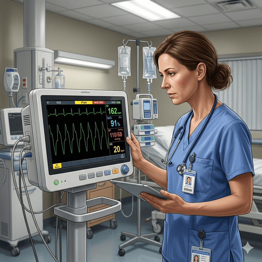
Cardiac rhythm interpretation is a critical skill in emergency and intensive care environments. One of the most challenging electrocardiogram (ECG) interpretations involves distinguishing ventricular tachycardia (VT) from supraventricular tachycardia with aberrancy (SVT with aberrancy). Both rhythms can appear as wide-complex tachycardias, which means the ECG shows a rapid heart rate with a widened QRS complex.
For the nurse and registered nurse (RN nurse) working in emergency departments, cardiac units, or intensive care settings, recognizing the difference between these rhythms is essential for patient safety. Incorrect interpretation may delay appropriate treatment or lead to dangerous interventions. Because of this importance, rhythm identification is commonly tested on the NCLEX and frequently discussed in advanced nursing education and many nursing bundle study resources.
This article explains the differences between ventricular tachycardia and supraventricular tachycardia with aberrancy, their ECG characteristics, and the role of nurses in identifying and managing these arrhythmias.
Understanding Wide-Complex Tachycardia
A wide-complex tachycardia occurs when the heart rate is typically greater than 100 beats per minute and the QRS complex is wider than 0.12 seconds on the ECG.
Wide-complex tachycardias usually fall into two main categories:
- Ventricular Tachycardia (VT)
- Supraventricular Tachycardia with Aberrant Conduction
Because these rhythms may appear similar on the ECG, careful analysis is required.
For the RN nurse, recognizing this difference can help guide urgent treatment decisions and support rapid communication with the healthcare team.
What Is Ventricular Tachycardia?
Ventricular tachycardia is a life-threatening arrhythmia that originates in the ventricles, the lower chambers of the heart.
During VT, abnormal electrical signals begin in the ventricles rather than the sinoatrial (SA) node. As a result, the heart beats rapidly and inefficiently.
Common causes include:
- Myocardial infarction
- Structural heart disease
- Electrolyte imbalances
- Drug toxicity
Because ventricular tachycardia can quickly deteriorate into ventricular fibrillation, it requires immediate medical attention.
The registered nurse must recognize this rhythm quickly and initiate appropriate emergency protocols.
What Is Supraventricular Tachycardia with Aberrancy?
Supraventricular tachycardia with aberrancy originates above the ventricles, usually in the atria or the atrioventricular (AV) node. However, abnormal conduction through the ventricles causes the QRS complex to appear widened.
This abnormal conduction often occurs due to:
- Bundle branch block
- Preexisting conduction pathway abnormalities
- Rapid heart rates affecting ventricular conduction
Although SVT with aberrancy can appear similar to ventricular tachycardia on the ECG, its origin is different and it may require different treatment strategies.
Therefore, accurate interpretation is extremely important in critical care nursing.
Key ECG Differences Between VT and SVT with Aberrancy
Distinguishing between these rhythms often requires careful ECG evaluation. Several features can help clinicians identify ventricular tachycardia.
AV Dissociation
One important clue is AV dissociation, where the atria and ventricles beat independently.
This feature strongly suggests ventricular tachycardia.
The RN nurse reviewing ECG strips should look for P waves that do not consistently relate to QRS complexes.
Capture Beats and Fusion Beats
Another sign of ventricular tachycardia is the presence of capture beats or fusion beats.
These occur when occasional normal sinus impulses briefly activate the ventricles during the tachycardia rhythm.
Although subtle, these findings strongly support a diagnosis of ventricular tachycardia.
QRS Complex Shape
The QRS morphology may also provide clues.
- VT often produces very wide and bizarre QRS complexes
- SVT with aberrancy usually resembles known bundle branch block patterns
Because these differences can be subtle, careful ECG interpretation is necessary.
Patient History and Clinical Context
Clinical history is another important factor.
For example:
- Patients with prior heart disease are more likely to develop ventricular tachycardia.
- Younger patients without structural heart disease may be more likely to experience SVT with aberrancy.
The nurse should always consider the patient’s clinical condition when evaluating arrhythmias.
Why Correct Identification Is Important
Misinterpreting ventricular tachycardia as supraventricular tachycardia can lead to inappropriate treatment.
For example, certain medications used to treat SVT may worsen ventricular tachycardia.
Because ventricular tachycardia can rapidly progress to cardiac arrest, it is often safer to assume wide-complex tachycardia is VT until proven otherwise.
This principle is commonly taught in advanced cardiac life support and critical care nursing education.
Nursing Responsibilities in Managing Tachycardia
The registered nurse plays a key role in recognizing and responding to abnormal cardiac rhythms.
Important nursing responsibilities include:
- Monitoring continuous ECG readings
- Recognizing abnormal rhythms quickly
- Assessing patient symptoms such as chest pain or dizziness
- Notifying the healthcare team immediately
- Preparing emergency medications or defibrillation equipment
Because rapid intervention may be necessary, the RN nurse must remain alert and prepared for sudden changes in patient condition.
Importance for NCLEX Preparation
Cardiac rhythm interpretation is a major topic in nursing education and appears frequently on the NCLEX exam.
Students preparing for the exam should understand:
- Differences between ventricular and supraventricular rhythms
- ECG characteristics of tachycardia
- Appropriate nursing responses to life-threatening arrhythmias
- Patient assessment priorities
Mastering these topics helps future registered nurses make safe clinical decisions in high-pressure situations.
How Nursing Study Bundles Support Learning
Many nursing students use structured study resources to review complex ECG topics.
A well-organized nursing bundle may include:
- ECG rhythm interpretation guides
- Visual rhythm comparison charts
- Step-by-step ECG analysis methods
- NCLEX-style practice questions
These resources help future RN nurses build confidence in recognizing life-threatening arrhythmias.
Final Thoughts
Differentiating ventricular tachycardia from supraventricular tachycardia with aberrancy is a critical skill in emergency and critical care medicine. Although both rhythms appear as wide-complex tachycardias, their underlying causes and treatment strategies can differ significantly.
Because rapid recognition is essential for patient survival, the nurse must carefully analyze ECG findings and consider the patient’s clinical condition. The registered nurse plays a vital role in monitoring cardiac rhythms, identifying abnormalities, and supporting rapid intervention.
For students preparing for the NCLEX, understanding these arrhythmias is an important part of advanced nursing education. With consistent practice and the support of structured resources such as a nursing bundle, future registered nurses can develop strong ECG interpretation skills and improve patient safety.