A Practical NCLEX Guide for the Nurse, Registered Nurse, and RN Nurse
Axis deviation is determined by evaluating QRS direction in leads I and aVF—and it can quickly signal ventricular hypertrophy, conduction blocks, or pulmonary disease.
For every nurse, registered nurse, and RN nurse, understanding ECG axis deviation is a core cardiac interpretation skill. Whether preparing for the NCLEX or working in critical care, telemetry, or emergency settings, axis interpretation strengthens clinical judgment and supports advanced nursing bundle cardiac assessment.
Let’s break it down in a way that makes sense clinically — not just academically.
What Is Cardiac Axis?
The cardiac axis represents the overall direction of ventricular depolarization in the frontal plane.
Think of it as the “electrical direction” the heart’s ventricles are firing.
Normal ventricular depolarization generally moves:
- Downward
- Leftward
- Toward the left ventricle (which has greater muscle mass)
Normal QRS Axis Range
NormalQRSAxis:−30°to+90°Normal QRS Axis: -30° to +90°NormalQRSAxis:−30°to+90°
If the electrical impulse falls within this range, the axis is considered normal.
For NCLEX purposes, remember:
- Normal axis = no major deviation
- Deviation suggests underlying pathology
The Quick Nursing Method: Lead I and aVF
The fastest way for a nurse to determine axis is by checking:
- Lead I
- Lead aVF
Step 1: Look at the QRS in Lead I
- Upright = positive
- Downward = negative
Step 2: Look at the QRS in Lead aVF
- Upright = positive
- Downward = negative
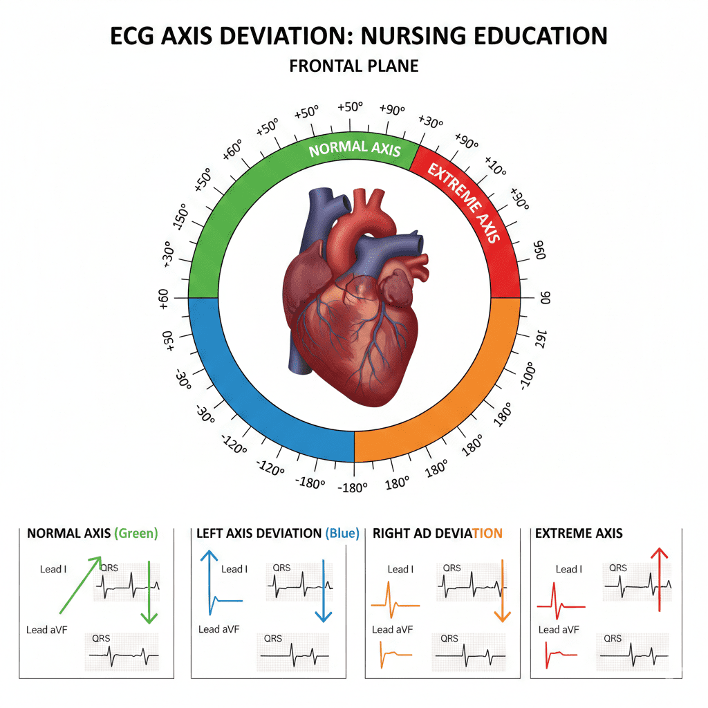
The 4 Axis Possibilities
1️⃣ Normal Axis
Lead I: Positive
aVF: Positive
→ Normal electrical direction
→ Common in healthy individuals
2️⃣ Left Axis Deviation (LAD)
LeftAxisDeviation:−30°to−90°Left Axis Deviation: -30° to -90°LeftAxisDeviation:−30°to−90°
Lead I: Positive
aVF: Negative
Clinical Meaning
Common causes:
- Left ventricular hypertrophy (LVH)
- Inferior myocardial infarction
- Left anterior fascicular block
- Chronic hypertension
For the registered nurse, this often correlates with:
- Long-standing hypertension
- Aortic stenosis
- Structural heart disease
LAD is a high-yield NCLEX cardiac concept.
3️⃣ Right Axis Deviation (RAD)
RightAxisDeviation:+90°to+180°Right Axis Deviation: +90° to +180°RightAxisDeviation:+90°to+180°
Lead I: Negative
aVF: Positive
Clinical Meaning
Common causes:
- Right ventricular hypertrophy (RVH)
- Pulmonary hypertension
- Pulmonary embolism
- Chronic lung disease (COPD)
For the ICU RN nurse, RAD may be an early clue of:
- Acute pulmonary embolism
- Severe hypoxia
- Right-sided heart strain
This is especially important in respiratory failure cases within a cardiac nursing bundle assessment.
4️⃣ Extreme Axis Deviation (“Northwest Axis”)
Lead I: Negative
aVF: Negative
Clinical Meaning
This is rare and often serious.
Associated with:
- Ventricular rhythms
- Severe conduction abnormalities
- Advanced cardiomyopathy
- Hyperkalemia
For the bedside nurse, this should trigger:
- Immediate rhythm evaluation
- Assessment of hemodynamic stability
- Electrolyte review
Why Axis Deviation Matters Clinically
Axis deviation is not just a number — it is a diagnostic clue.
It can indicate:
- Chamber enlargement
- Conduction blocks
- Acute pulmonary pathology
- Electrolyte disturbances
- Structural heart disease
In a structured nursing bundle, ECG axis interpretation complements:
- Vital sign trends
- Oxygenation status
- Electrolyte labs
- Cardiac biomarkers
NCLEX-Focused Nursing Pearls
✔️ Left axis deviation → Think LVH or inferior MI
✔️ Right axis deviation → Think pulmonary cause
✔️ Extreme axis → Think ventricular rhythm or severe conduction issue
✔️ Always interpret axis with the full clinical picture
For the RN nurse preparing for the NCLEX, exam questions often present:
- An ECG strip
- A patient with hypertension or COPD
- A question asking which chamber is enlarged
Axis interpretation often gives the answer.
Advanced Clinical Integration for the Registered Nurse
Axis deviation becomes especially powerful when combined with:
- QRS duration analysis
- ST-segment changes
- Bundle branch blocks
- Electrolyte abnormalities (like hyperkalemia)
For example:
• Left axis + wide QRS → possible left bundle branch block
• Right axis + hypoxia → possible pulmonary embolism
An experienced nurse doesn’t just identify the axis — they connect it to the patient.
Step-by-Step Nursing Bundle for ECG Axis Assessment
- Assess patient stability first
- Evaluate rhythm and rate
- Check QRS width
- Determine axis (Lead I + aVF)
- Correlate with clinical findings
This systematic approach strengthens critical thinking and elevates nursing practice from task-based to analytical.
Final Thoughts for the Nurse and RN Nurse
Axis deviation is one of the simplest yet most powerful ECG tools available to the registered nurse.
It provides:
- Early diagnostic clues
- Insight into cardiac remodeling
- Indicators of pulmonary stress
- Guidance for urgent escalation
For NCLEX success and real-world cardiac care, mastering axis interpretation builds confidence and clinical precision.
When you see an ECG, don’t just look at the rhythm.
Look at the direction.
That direction tells a story.