Electrocardiograms (EKGs) are one of the most tested skills on the NCLEX and a critical responsibility for every nurse and registered nurse (RN nurse) at the bedside. Understanding the 12-lead EKG and which lead corresponds to which part of the heart helps in recognizing ischemia, infarction, and arrhythmias early. For nursing students and graduates, this knowledge can feel overwhelming—but when broken down by regions, it becomes simple and manageable.
This guide simplifies 12-lead EKG interpretation and highlights what every nursing bundle should include for exam prep and clinical practice.
🔹 Why Nurses Must Master 12-Lead EKGs
- NCLEX Prep: Questions on EKG interpretation often test whether the nurse can identify which area of the heart is affected.
- Patient Safety: Rapid recognition of ST-elevation, depression, or arrhythmias can save lives.
- Clinical Confidence: A registered nurse who can read 12-leads accurately becomes a valuable member of the healthcare team.
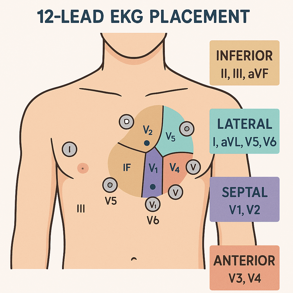
🔹 The 12 Leads and Heart Anatomy
A 12-lead EKG provides 12 different “views” of the heart’s electrical activity. Each set of leads looks at a specific anatomical region:
1. Inferior Leads – II, III, aVF
- Sees: Inferior wall of the heart
- Artery Involved: Right coronary artery (RCA)
- Nursing note: Watch for bradycardia and AV blocks with RCA infarctions.
2. Lateral Leads – I, aVL, V5, V6
- Sees: Lateral wall of the left ventricle
- Artery Involved: Left circumflex artery (LCX)
- Nursing note: NCLEX loves to test this region for left-sided ischemia.
3. Septal Leads – V1, V2
- Sees: Interventricular septum
- Artery Involved: Left anterior descending artery (LAD)
- Nursing note: Bundle branch blocks often show up here.
4. Anterior Leads – V3, V4
- Sees: Anterior wall of the left ventricle
- Artery Involved: Left anterior descending artery (LAD)
- Nursing note: Anterior infarctions have high mortality—recognition is critical.
🔹 Quick Memory Trick (NCLEX Friendly)
👉 “I See All”
- I, aVL → Lateral
- II, III, aVF → Inferior
- Septal → V1, V2
- Anterior → V3, V4
- Lateral again → V5, V6
This mnemonic helps nursing students recall which leads correspond to which areas during NCLEX exams.
🔹 Nursing Priorities with Abnormal 12-Lead Findings
- ST Elevation – Possible MI → Call rapid response/code, prepare for cath lab.
- ST Depression or T-Wave Inversion – Possible ischemia → Give O2, nitrates, monitor.
- Arrhythmias – Ventricular or atrial arrhythmias → Follow ACLS protocol, monitor closely.
- Bundle Branch Blocks – Assess stability, monitor for progression.
💡 Nurse Tip: Always compare with previous EKGs if available.
🔹 The Nurse’s Role in 12-Lead EKGs
- Place leads correctly (misplacement = misdiagnosis).
- Recognize abnormal changes early.
- Communicate findings promptly to the healthcare team.
- Use nursing bundles or reference guides to reinforce learning and NCLEX prep.
✅ Final Thoughts
Mastering 12-lead EKG interpretation is essential for every nurse and registered nurse preparing for the NCLEX. By simplifying leads into regions—inferior, lateral, septal, and anterior—you can quickly identify which part of the heart is affected and respond appropriately. Whether in exams or real-life emergencies, this skill can save lives.
❓ FAQ: 12-Lead EKG Simplified
A 12-lead EKG provides a complete view of the heart’s electrical activity from different angles. It helps nurses and registered nurses identify ischemia, infarction, arrhythmias, and conduction blocks, making it a key skill tested on the NCLEX.
The anterior leads are V3 and V4, which visualize the front wall of the left ventricle supplied by the left anterior descending (LAD) artery.
Leads II, III, and aVF reflect the inferior wall of the heart, usually supplied by the right coronary artery (RCA).
The lateral leads are I, aVL, V5, and V6. They assess the lateral wall of the left ventricle, which is often affected in circumflex artery blockages.
For nurses and RN nurses, recognizing which lead corresponds to which heart region helps in early detection of myocardial infarction (MI), prioritization in emergencies, and success on NCLEX-style questions.
A common nursing bundle tip is to think of the chest leads (V1–V6) moving from the septum → anterior → lateral wall, while limb leads (I, II, III, aVR, aVL, aVF) provide a “global” view of the heart.