Electrolytes play a vital role in the heart’s electrical system. Even small shifts in potassium, calcium, magnesium, or sodium levels can show up on an EKG. Recognizing these changes can help nurses act quickly and prevent serious cardiac complications.
Let’s break it down by each electrolyte imbalance and what to watch for on the monitor.
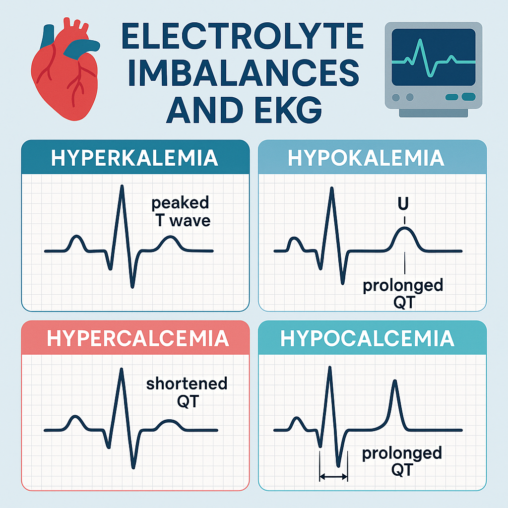
🧪 1. Potassium: The EKG Game-Changer
Hyperkalemia (High Potassium)
- Causes: Kidney failure, potassium-sparing diuretics, tissue damage (burns, trauma).
- EKG Signs:
- Peaked T waves (early sign)
- Widened QRS complex
- Flattened or absent P wave
- Sine wave pattern (late and dangerous)
- Why It Matters: Hyperkalemia can lead to life-threatening arrhythmias like ventricular fibrillation or asystole.
Hypokalemia (Low Potassium)
- Causes: Diuretics, vomiting, diarrhea, poor intake.
- EKG Signs:
- Flattened T waves
- U waves (extra wave after the T)
- ST depression
- Prolonged QT interval
- Why It Matters: Can cause deadly rhythms like torsades de pointes and ventricular tachycardia.
💀 2. Calcium: The Muscle Conductor
Hypercalcemia (High Calcium)
- Causes: Hyperparathyroidism, cancer, excess vitamin D.
- EKG Signs:
- Shortened QT interval
- Possible heart block in severe cases
- Impact: Increased risk of arrhythmias but often silent unless very high.
Hypocalcemia (Low Calcium)
- Causes: Hypoparathyroidism, kidney failure, pancreatitis.
- EKG Signs:
- Prolonged QT interval
- T wave changes
- Why It Matters: High risk for torsades de pointes and seizures.
⚡ 3. Magnesium: The Stabilizer
Hypermagnesemia (High Magnesium)
- Causes: Kidney disease, magnesium-containing antacids or IV overuse.
- EKG Signs:
- Prolonged PR interval
- Widened QRS
- Bradycardia
- Risk: Can lead to complete heart block or asystole if untreated.
Hypomagnesemia (Low Magnesium)
- Causes: Chronic alcoholism, poor diet, diarrhea, diuretics.
- EKG Signs:
- Prolonged QT interval
- Torsades de pointes
- Flattened T waves
- Why It Matters: Often seen with low potassium; correcting magnesium may fix arrhythmias when potassium alone doesn’t.
💧 4. Sodium: The Neurologic Marker
While sodium imbalances (hypernatremia or hyponatremia) primarily affect the brain, extreme cases can indirectly influence the heart through fluid shifts, seizures, or changes in mental status leading to injury. EKG changes are not usually direct, but nurses should be alert for secondary cardiac impacts.
🧠 Quick Memory Aids:
- Potassium Peaked = Peaked T waves
- Low Potassium = U wave
- High Calcium = Short QT
- Low Calcium or Magnesium = Prolonged QT
- Low Magnesium = Think Torsades
🩺 Nurse Tips: What to Do
- Always correlate lab values with EKG readings.
- Notify the provider immediately for serious findings like:
- Peaked T waves in hyperkalemia
- Prolonged QT in hypocalcemia or hypomagnesemia
- U waves in hypokalemia
- Monitor closely for changes in vitals, rhythm, and patient status.
- Be ready to administer replacement therapy or emergency interventions like calcium gluconate or insulin/dextrose in hyperkalemia.
📋 Final Thoughts
Reading EKG changes linked to electrolyte imbalances is a must-have skill for every nurse. Understanding the “why” behind these patterns helps you act fast and improve patient safety. When in doubt, double-check the labs, trust your monitor, and communicate with your team.